For the first time, a scoring scale has been developed to determine the degree of cardiac dysfunction in congenital heart defects using Doppler ultrasonography of the pulmonary artery and vein, ejection fraction and stroke volume of the ventricles of the fetal heart in the prenatal period;
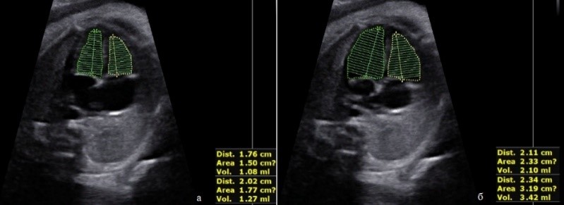
Evaluation of ventricular EF. a) ventricular ESV; b) ventricular EDV;
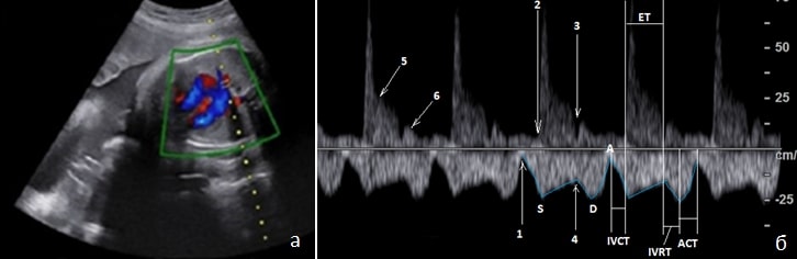
When using color Doppler imaging, both the pulmonary vein and the pulmonary artery are included in the control volume. Doppler spectrum of blood flow in the pulmonary artery (above the baseline) and pulmonary vein (below the baseline). 1 - atrioventricular valve closure point, 2 - pulmonary valve opening point, 3 - pulmonary valve closure point, 4 - atrioventricular valve opening point, 5 - dicrotic notch, 6 - wave caused by vascular wall turgor. ET - ejection time, IVCT - isovolumetric contraction time, IVRT - isovolumetric relaxation time, ACT - atrial contraction time. For the first time, the presence of four types of anatomical variants of the fetal portal venous system structure has been proven based on ultrasound examination data - T-, X-, H-shaped types and in the form of trifurcation in the diagnosis of fetal venous system development anomalies;
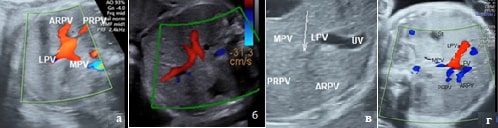
Connection with the main portal vein and its branches a) T-shaped type I; b) X-shaped connection type II; c) H-shaped type III, the connecting vessel is indicated by an arrow, d) a variant in the form of a trifurcation. In the diagnostics of abnormal pulmonary venous return, new Dopplerographic signs have been proven, such as "connection of all four pulmonary veins at one point (X-shaped type of connection)" in the supracardiac type of TADPV and "a sign of the mutual arrangement of the descending vertical vein with the aorta and descending vein" in the infracardiac type;
Supracardial type of TADLV. a) section at the level of confluence of the pulmonary veins, sign "X"; b) cross-section at the level of the main vessels and vertical vein;
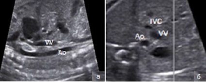
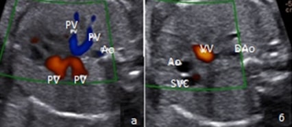
Infracardiac type of TADLV. a) relative position of the aorta and vertical vein in the sagittal section of the chest; b) relative position of the aorta, inferior vena cava and vertical vein in the abdominal cavity
In order to improve the timely and accurate diagnosis of congenital heart defects in the prenatal period, the protocol of extended fetal echocardiography has been improved using additional eleven heart sections and Doppler evaluation of intracardiac hemodynamics;
In women at risk, in order to reduce perinatal mortality and disability rates in children, ultrasound diagnostics of congenital heart defects in the prenatal period has been improved by using a scheme of triple screening ultrasound examination of the heart and extended fetal echocardiography.
The necessity of using ADF and B-Flow modes in patients with carotid artery atherosclerosis has been proven for a more accurate assessment of the degree of stenosis, the contour of the atherosclerotic plaque due to the absence of the flow dependence angle and the possibility of detecting low-speed flows;
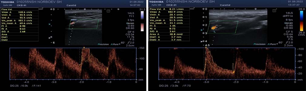
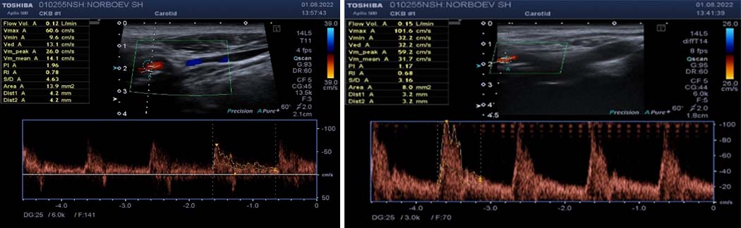
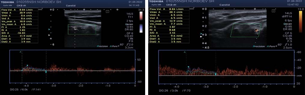
Standard values of total cerebral blood flow by age groups have been developed to characterize the features of carotid atherosclerosis in the Uzbek population;
To improve the accuracy of assessing the volumetric cerebral blood flow in carotid artery atherosclerosis, a regression model has been developed using the cerebrovascular reserve index;
It has been proven that a decrease in the cerebrovascular reserve index below 0.8 according to ultrasound Dopplerography in patients with a stenosis degree above 50% is a sign of hemodynamically significant arterial damage associated with a decrease in the volume of cerebral blood flow.
Right ICA, flow volume 360 ml/min - Left ICA, flow volume 210 ml/min.
Right ECA, flow volume 120 ml/min - Left ECA, flow volume 150 ml/min.
Right PA, flow volume 40 ml/min - Left PA, flow volume 40 ml/min.
Example of assessment of the CBC in an asymptomatic 67-year-old patient with significant stenosis of the right ICA (60%) with values of volumetric cerebral blood flow within the reference values (920 ml).
On November 30, 2024, the Department of Ultrasound Diagnostics-1 of the CDPQMW, in cooperation with "SAMSUNG MEDISON", held a scientific and practical seminar with international participation on the topic "Advanced Course: Ultrasound Research and Using Artificial Intelligence in the Practice of an Obstetrician-Gynecologist."
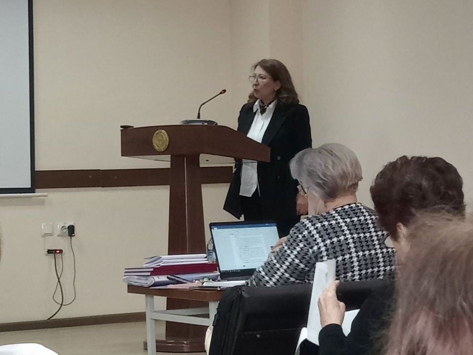
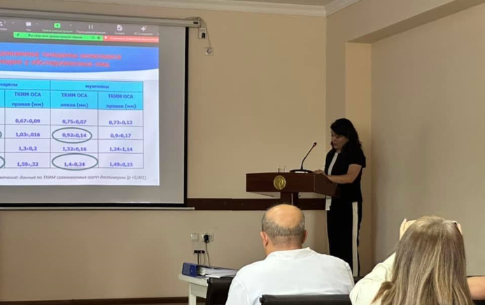
On November 21, 2024, at the Scientific Council for the Awarding of Academic Degrees DSc.04/30.12.2019.tib.77.01 at the Republican Specialized Scientific and Practical Medical Center of Oncology and Radiology, the Head of the Department, Normuradova N.M., successfully defended her dissertation for the degree of DSc in Clinical Radiology 14.00.19 "Strategy and tactics of ultrasound diagnostics of congenital fetal heart defects."
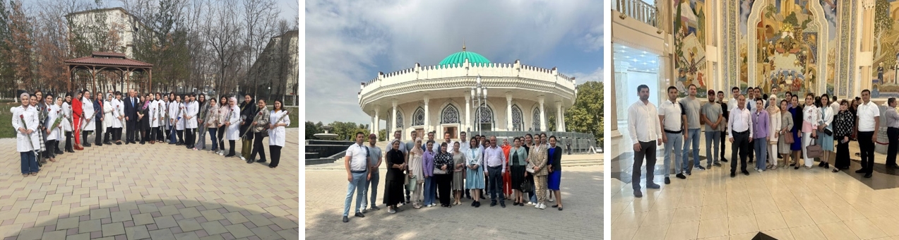
Department staff and students actively participate in spiritual and educational, sports events held by the Ministry of Health of the Republic of Uzbekistan and the Center for the Development of Professional Qualification of Medical Workers.
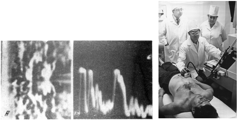
It has been 63 years since Professor Fozilov Akram Akmalovich became one of the first in the world to receive two- and one-dimensional ultrasound images of breast cancer in Uzbekistan. November 3, 1961.
On June 14, 2024, our department's assistant Ikramova Z.T. successfully defended her scientific work on the topic "Comprehensive assessment of Dopplerographic indicators of general cerebral blood flow in carotid atherosclerosis" for the degree of PhD 14.00.19 - Clinical Radiology.
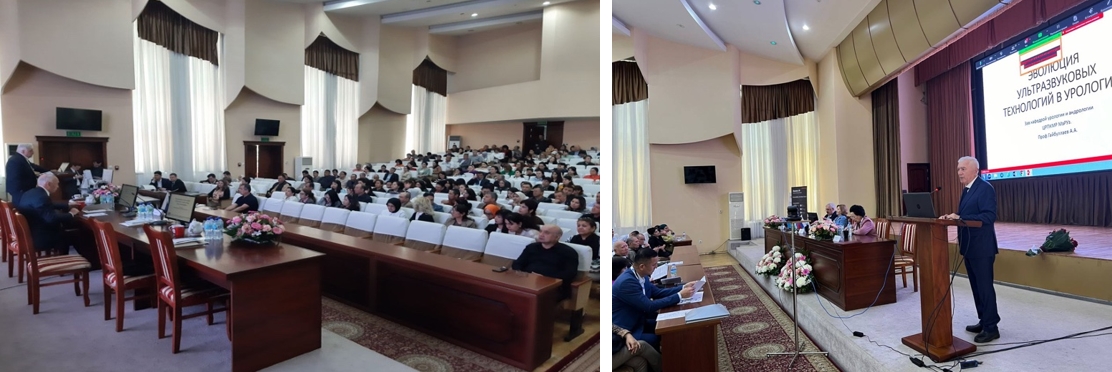
On April 18-20, a scientific and practical conference was held with the international participation "Innovative technologies of ultrasound diagnostics in medicine," organized by the department with the support of the Ministry of Health and CDPQMW. Along with Uzbek specialists, scientists from Kazakhstan, Russia, and Kyrgyzstan participated with presentations. The conference was also attended by about 400 ultrasound doctors, clinical residents, and magistrates.
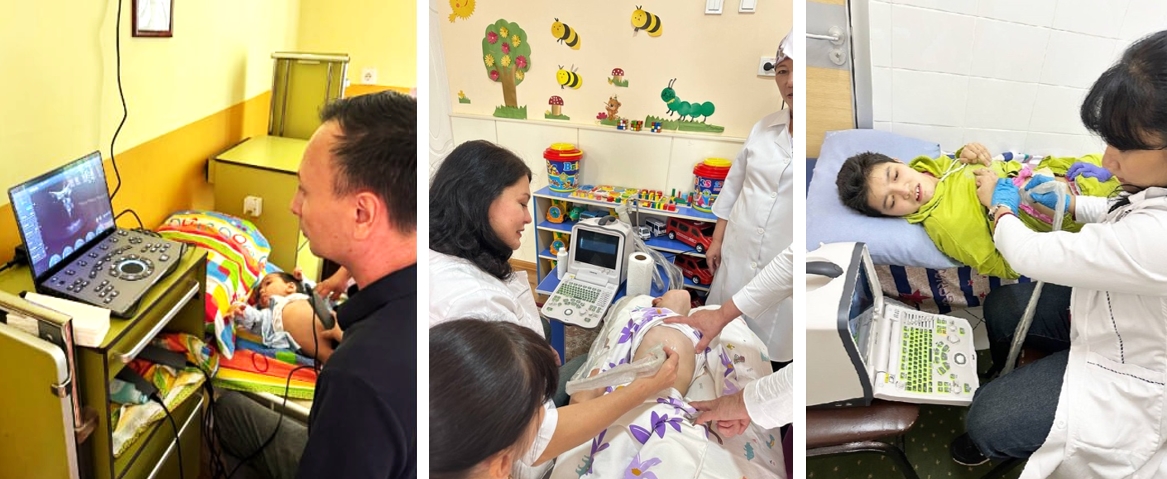
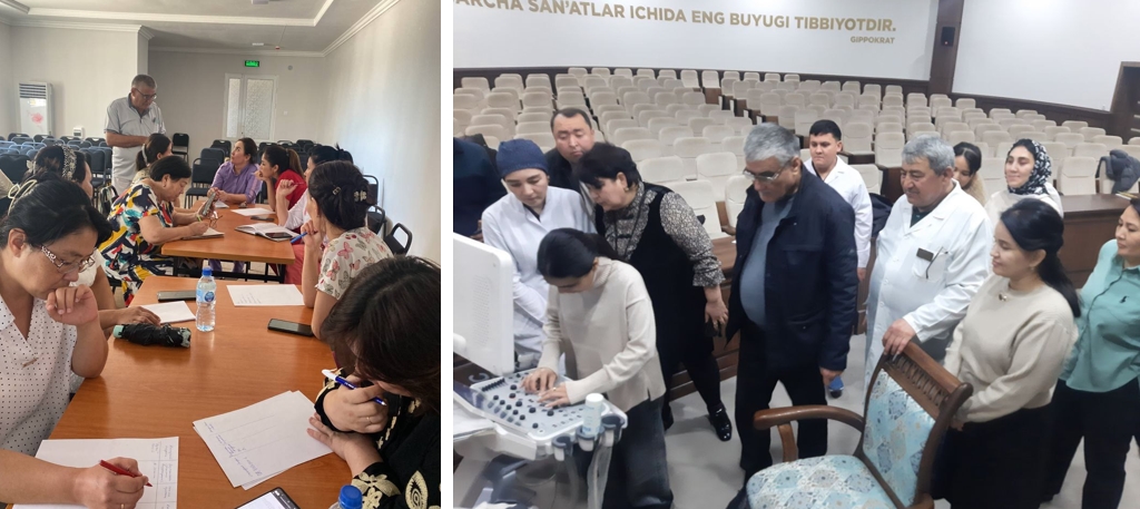
At the specialized home for children with psychophysical developmental disorders in Tashkent region, our department staff participated in an in-depth medical examination of the children's home's residents with qualified specialists from the Center for Professional Development of Medical Workers of the Ministry of Health.
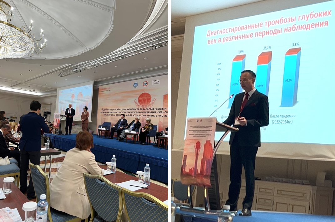
С On April 12-13, 2024, department staff, along with specialists from Austria, Russia, UAE, and Korea, participated and delivered presentations at the International Scientific and Practical Conference "Women's Health in Radiology" of the Radiology Association held in Astana, Kazakhstan.
On January 26, 2024, at the Department of USD-1 of the CDPQMW, Associate Professor of the Department of Obstetric Gynecology of the N.I. Pirogov Russian National Medical University, Irina Vasilievna Karachenseva conducted a seminar-master class on the topic "Opportunities of Ultrasound Diagnostics in Pediatric Gynecology." Professor of the Department Fazilov A.A., Head of the Department Normuradova N.M., Associate Professor of the Department Rasulova M.M. and other department staff discussed the problems of diagnostics in pediatric gynecology with a Russian specialist.
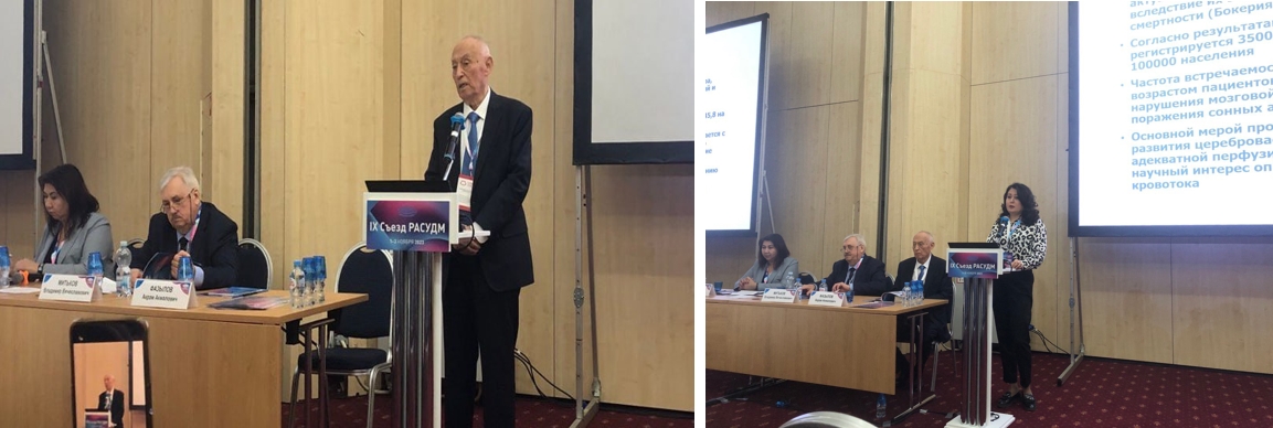
On November 1-3 of this year, a scientific session on the topic "Russia Meets Uzbekistan" was held at the 9th Congress of the RASUDM. 9 reports of USD specialists from Uzbekistan were heard (including 4 from the USD-1 department). The department is a co-author of this scientific session.
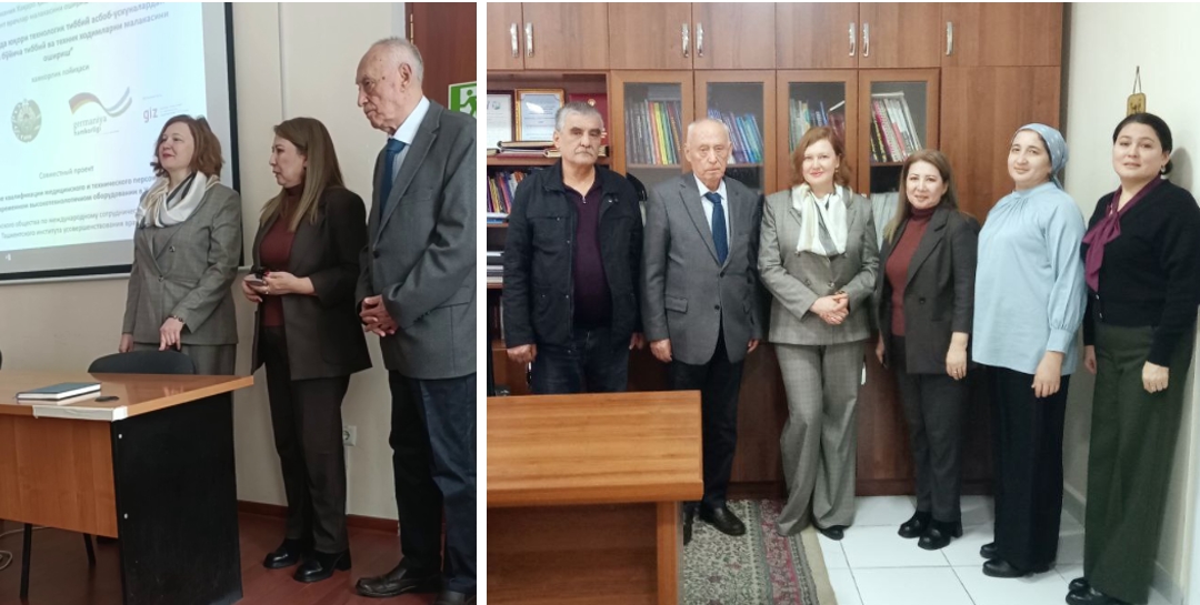
Within the framework of the "Management of Advanced Medical Technologies in Uzbekistan" project, conducted by GIZ and GOPA (German Society for International Cooperation), department staff were engaged as international trainers and experts to conduct training sessions on "Care and Maintenance of Ultrasound Devices by Medical Workers" in 6 experimental regions (Republic of Karakalpakstan, Andijan, Fergana, Namangan, Samarkand, Tashkent city). 122 specialists from the Centers for Perinatal Diagnostics and Emergency Medical Care participated in the training and underwent certification.
On April 28-29, 2023, the department, with the support of the Ministry of Health of the Republic of Uzbekistan, the Republican Specialized Scientific and Practical Medical Center, and company Samsung, held a scientific and practical conference with international participation on "Current issues of prenatal neurosonography and echocardiography." It was attended by about 300 specialists in ultrasound diagnostics, guests from Russia, Belarus, the Republic of Korea.
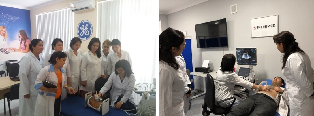
Practical classes in simulation centers at the department are continuing at a high pace. These centers have phantoms for training in the following areas: fetal, abdominal, neurosonography, breast, gynecology, as well as 4 modern ultrasound devices that meet all the requirements.
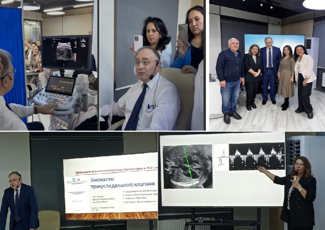
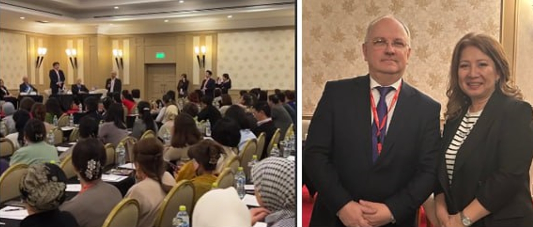
On February 15, 2025, the 1st Department of Ultrasound Diagnostics at the CFPQMW conducted a seminar with international participation on the topic "Innovative Ultrasound Technologies in Maternal and Child Health Protection." Lectures and master classes were held with the participation of Professor of the Department of Internal Diseases of the Institute of Medical Education within the Federal State Budgetary Educational Institution "Yaroslav the Wise Novgorod State University" (Great Novgorod), Head of the Ultrasound Diagnostics Department of the "Regional Clinical Hospital" of the State Budgetary Healthcare Institution of the Vladimir Region (Vladimir), Executive Secretary of the Russian Association of Ultrasound Diagnostics Specialists in Medicine (RASUDM) (Moscow), Official Lecturer of RASUDM Bulanov Mikhail Nikolaevich, as well as employees of the Department of Ultrasound Diagnostics of the CFPQMW. 157 ultrasound doctors from various prenatal screening rooms participated in the event.